- Category: Volume 12
- Hits: 8262
Study of the genetic diversity of the local cattle population in Tunisia by the AFLP method
| [Volume 12, Article 4] | 4961 kB |
R. Baccouche 1
S. Bedhiaf 1
B. Jemmali 2
1 Institut National Agronomique de Tunis.
2 Laboratoire ADIPARA, Département de la Production Animale, Ecole Supérieure d’Agriculture Mateur, Tunisie.
Abstract - The survey was conducted in North West Tunisia on 154 native cattle owners. Blood samples were collected so that genetic diversity can be studied. The study was based on 2 primers combinations. Amplified fragment length polymorphism (AFLP) was used to detect within and among population genetic diversity.
The polymorphism information content (PIC) was 0.31. The differentiation coefficient between populations (Gst) was 0.06. These results point toward that the main genetic variation was within the population (94%) and the remaining (6%) of the variation was among populations. The gene flow (Nm) was relatively high (7.56). These results show the dangerous situation in which native cattle are encountered.
Keywords: native cattle, population, AFLP, genetic polymorphism.
-
Introduction
The local cattle population in Tunisia is formed by two breeds “Blonde du cap bon” and “Brune de l’Atlas” was clustered into two branches based on the gray and the fawn one. This population is characterized by its hardiness (Atti et al., 1972). Besides, it constitutes a very important socio-economic element and contributes largely to food in rural areas. This population has gathered ability to adapt to the environment, food availability, the pathological contexts and modes of operation.
The morphological traits are greatly influenced by environment, thus diversity studies based on morphological traits are not reliable (Maryam M et al., 2010). DNA markers made possible a fast survey of genetic diversity and genetic structure (Xue et al., 2006). AFLP (Amplification fragment length polymorphism) is a DNA fingerprinting technique, first described by Vos et al. (1995). It is based on selective PCR amplification of DNA restriction fragments under stringent conditions (Vos et al., 1995). Having the potential to generate a large number of polymorphic loci (Powell et al., 1996), comparative studies indicated that AFLP is a powerful tool to detect levels of genetic diversity and genetic structure of natural populations in many animals’ species. The advantages of this method are reproducibility, high level of polymorphism detection genome wide distribution of markers and no pre-requisite of knowledge of the genome being studied (Muller and Wolfenbarger, 1999). Few studies have aborted the evaluation of genetic diversity of animals mostly cattle using AFLP technique. For this reason, this study approach to describe the different steps of this technique.
To show interest to the genetic diversity of the indigenous cattle population in Tunisia, the objective of this study was to investigate genetic diversity and population genetic structure of 154 cattle genotypes of North regions of Tunisia belonging to 3 regions (Bizerte, Béja and Jendouba) using AFLP technique.
-
Materials and methods
-
Sample collection
-
A total of 154 samples of native cattle were collected from eight locations as shown in figure 1 (Bizerte: Séjnène, Shiri; Béja: Nevza, Chottezoueraa, Loulija ; Jendouba: Ghareddimaou, Tabarka and Ain Drahem). Venous blood was taken from each individual’s from the jugular vein and mixed with anticoagulant (EDTA) and then kept at -20°C for subsequent DNA extraction.
|
|
|
Figure 1. The map of the locations of samples collected of native bovine population
|

Groups of animals were performed according to their geographical location. Only the “Blonde du Cap Bon” was a group by itself in three governorates because it is very limited (only seven individuals in three governorates). Acronyms have been affected; BAB: individual of “Brune de l’Atlas” in Béja, BAZ: individual of “Brune de l’Atlas” in Bizerte, BAJ: individual of “Brune de l’Atlas” in Jendouba , BC: Blonde du Cap Bon in 3 governorates.
|
Table 1. Summary of all samples in this study |
|
|
Governorate |
Number of samples |
|
Bizerte |
35 |
|
Béja |
50 |
|
Jendouba Total |
69 154 |
-
DNA extraction
Genomic DNA extraction was extracted from blood samples according to standard phenol and chloroform method. The DNA concentration was estimated spectrophotometrically and its integrity was checked by analytical [0.8% (W/V)] agarose gel electrophoresis (Sambrook et al., 1989).
-
AFLP analysis
The AFLP process was carried out using the protocol of Vos et al. (1995). The principle of this technique is based on three phase’s namely successive digestion-ligation, pre-amplification and selective amplification.
-
Digestion-ligation
-
First enzymatic digestion
-
This digestion is carried out with the enzyme EcoRI to cut uncommon and restriction sites of 6 bases (Table 2). Genomic DNA (2µl) with a concentration equal to 500ng is diluted in 8µl of ddH2O. The reaction mixture contains 5 U of EcoRI, 1x of buffer and ddH2O. The total volume is 20µl was vortexed and placed in a water bath at 37°C during 4h, maintained at a temperature of -20°C.
|
Table 2. Restriction enzymes used for AFLP |
||
|
Enzyme |
Sequence recognized |
Cleavage site |
|
5'---G AATTC---3' |
||
|
Tru1I(MseI) |
5'---T TAA---3' |
|
-
Second digestion
The product of the first digestion underwent a second digestion with 5U of MseI in a total volume of 40µl (Table 3) during 4 hours in a water bath at 65°C. The restriction enzymes were finally inactivated by incubation of the samples for 10min at 85°C.
|
Table 3. Reaction mixture composition for the enzymatic digestion MseI |
|||
|
Composition of mixed |
Concentration of stock solution |
Final concentration |
Volume required (µl) |
|
ddH2O |
- |
- |
17,5µl |
|
Buffer (Fermentas, Life Science) |
10x |
1x |
2 |
|
MseI (Fermentas, Life Science) |
10U/µl |
5U |
0,5 |
|
DNA |
- |
500ng |
20 |
|
Total volume |
- |
- |
40 |
Then, digested DNA was checked for its quality and quantity fragment by 1.5% agarose gel electrophoresis. A 1.5% agarose gel was prepared (1.5 g of agarose in 100 ml of TBE). 5µl of the digested product was added to the wells. Finally, the migration was performed during 30min at 130 volts.
-
Ligation
Adapters were prepared by proceeding to the mixture of equimolar amounts of the two oligonucleotides single strand for a final concentration of 5 pmol/µl of EcoRI and 50 pmol/µl of Tru1I. The oligonucleotides were hybridized by incubation at 95°C for 5min, then at 65°C during 15min, followed by a gradual return to room temperature. For the ligation, a volume of 10µl of the reaction mixture (Table 4) containing specific adapters for both endonucleases and ligase was added to the volume of 20µl of the digested DNA.
|
Table 4. List of adapters used for ligation |
|
|
Adapter-EcoRI (Adaptateur-E) |
Adapter-MseI (Adaptateur-M) |
|
F : 5’CTC GTA GAC TGC GTA CC 3’ R: 5’AAT TGG TAC GCA GTC TAC3’ |
F : 5’ GAC GAT GAC TCC TGA G 3’ R: 5’ TAC TCA GGA CTC AT 3’ |
-
DNA amplification
The amplification of the product digested and ligated occurs in two phases.
-
Pre-amplification
In this case, the nucleotide A was chosen for the 3’ end of the EcoRI and the nucleotide C to the Tru1I. Dilution of the product digested and ligated to 1:5 in buffer was performed. Then we took 5 pmol/µl of adapter E (Applied Biosystem), 50 pmol/µl of adapter M, 1 U of T4DNA, 1x of buffer and we add ddH2O. Afterwards, we added a reaction mixture which contains 1x of buffer (biomatik), 1.5 mM of MgCl2, 0.2 mM of dNTP, 50 ng of EcoRI-A, 50 ng of MseI-C and 1U of Taq polymerase.
Pre-selective amplification was performed in a thermocycler (C1000TM, Bio-Rad) with the following program: pre-denaturation at 94°C during 2min; 30 cycles: at 94°C during 30s for denaturing, at 56°C for 30s for annealing and at 72°C for 1min for the elongation. These cycles were completed by a final extension during 10min at 72°C.
-
Selective amplification
The specific and selective combinations of primers are: EcoRI-AGG/MseI-CTT; EcoRI-ACT/MseI-CTT. PCR products contain a fluorescent dye, when they pass to the sequencer for their identification. A dilution of 1:20 was made in TE of amplified products. We took 5µl of diluted product and it was added 20µl of reaction mixture (table 5). The total volume of selective amplification was 25µl. The amplification was performed in a thermocycler with the following program: pre-denaturation at 94°C during 2min; 10 cycles: at 94°C during 20s for denaturing, at 66°C for 30s for annealing and at 72°C for 2min for the elongation. And 20 cycles: at 94°C during 20s for denaturing, at 56°C for 30s for annealing and at 72°C for 2min for the elongation. These cycles were completed by a final extension during 2 min at 72°C.
|
Table 5. Composition of the reaction mixture for the selective amplification |
||||
|
Reagents |
Concentration of stock solution |
Final concentration |
||
|
dH2O Gibco |
--- |
--- |
||
|
Tampon |
10x |
1x |
||
|
MgCl2 Dntp |
25mM 25µM |
1.5mM 0.2mM |
||
|
EcoRI-AXX |
10µM |
5ng |
||
|
MseI-CYY |
10µM |
30ng |
||
|
Taq polymerase |
5U/µl |
1U/µl |
||
|
Total volume |
--- |
--- |
||
-
Revelation of the amplification products
The selective amplification products were prepared as a mix of the following: 1.0µl of each fluorescently labelled PCR products, 12.0µl of formamide and 0.5µl of Gene Scan TM size standard (Applied Biosystem). The mixed products were then separated on an ABI PRISM 3130 Genetic Analyser (Applied Biosystem) sequencer in the Laboratory of Animal Biotechnology (National Gene Bank of Tunisia) using a polymer 3130 POP7TM (Applied Biosystems). Data were collected automatically and analysed using software GeneMapper 4.0 (Applied Biosystem).
-
Data statistic and analysis method
By AFLP polymorphic bands statistical method, each allele was scored as present (1) or absent (0) for each loci. Each band was scored which is clear and repeated, and the size ranged from 100-500pb in length. The polymorphism information content (PIC) of each marker was calculated using PIC=2fi (1-fi); were fi is the frequency of the marker fragments which were present (Roldan-Ruiz et al., 2000). Resolving power (Rp) of each combination of primers was calculated using the formula: Rp= Ib were Ib= 1-[2(0.5-p)], and p is the proportion of the individual containing the I band (Prevost and Wilkinson, 1999). Matrix of similarity between individuals was obtained by NTSYS pc softwarepackage, version 2.02 (Rohlf, 1998), using Dice Similarity Index and Nei and Li coefficient (Nei and Li, 1979). The cluster analysis was performed using the unweighted pair group method of arithmetic averages (UPGMA) procedure (Sokal and Michener, 1958). Some genetic diversity parameters in populations i.e. percentage of polymorphic band (PPB), mean number of alleles per locus (Na), mean effective number of alleles per locus (Ne), Nei’s genetic diversity (H) (Nei, 1973), Shannon’s Index (I) (Lewontin, 1972), gene flow [Nm= 0.5 (1-Gst)/Gst] (McDermott and McDonald, 1993), Nei’s genetic distance (Nei, 1978), coefficient of gene differentiation between populations (Gst) were calculated by POPGENE 1.32 software (Yeh et al., 1999).
3. Results and discussions
3.1. Evaluation combinations of primer
The two AFLP primer produced a total of 314 scorable bands for the 154 individuals across four populations, of which 247 (78.66%) are polymorphic. Resolving power (Rp) varied from 56.7 to 69.5 and the polymorphism information content (PIC) ranged from 0.31 to 0.32 (Table 6).
|
Table 6. Polymorphic bands and resolving power generated by the two combination of primer used in the inter specific analysis |
|||||
|
Primer combination |
NTB |
NBP |
PBP |
Rp |
PIC |
|
E-AGG/M-CTT |
171 |
134 |
78.36 |
69.5 |
0.32 |
|
E-ACT/M-CTT |
143 |
113 |
79.02 |
56.7 |
0.31 |
|
Total |
314 |
247 |
- |
126.2 |
0.32 |
Different electrophoresis profiles obtained consist of 113 to 134 bands respectively for combination of primer E-ACT/M-CTT and E-AGG/M-CTT, with an average equal to 123 (Table 6). After calculating PIC for each population in each combination, and those for the two combinations merged, it turns out that the highest value (0.34) is that of BAJ (Table 7).
|
Table 7. PIC values for each populations |
|||
|
Population |
E-AGG/M-CTT |
E-ACT/M-CTT |
Two combinations |
|
BAZ |
0.35 |
0.33 |
0.35 |
|
BAB |
0.29 |
0.30 |
0.29 |
|
BAJ |
0.34 |
0.33 |
0.34 |
|
BC |
0.30 |
0.30 |
0.30 |
These results suggest the effectiveness of AFLP primer combinations used and confirm that the AFLP technique generates a large number of polymorphic markers (Morgante and Olivier., 1993). This is confirmed by the value of Rp obtained. The combination of primer E-AGG/M-CTT presents the highest value of Rp, PIC and PPB, thus contributing to better characterization of native cattle (Table 6).
3.2. Genetic variations within populations
Estimates of genetic diversity in each population are summarized in Table (8), the percentage of polymorphic bands (PPB) at the population level ranged from 83.81% in BC population to 100% in BAJ population. The observed number of alleles (Na) ranged from 1.84 in BC population to 2 in BAJ population. The effective number of alleles (Ne) were from 1.51 to 1.58 and the Shannon information index (I) ranged from 0.44 to 0.51, mean Nei’s gene diversity was varying from 0.29 (BC population) to 0.34 (BAJ population), total diversity (Ht) was 0.34. The average of diversity among populations (Hs) was 0.32 (Table 9). The highest percentage of polymorphic loci, gene diversity, Shannon information index, the observed number of alleles (Ne) was observed in BAJ population and the lowest was observed in BC population.
The result Gst=.06 shows significant differentiation between populations in allele frequencies. We can therefore note that a little percentage of genetic diversity is distributed among populations or ecotypes.
|
Table 8. Statistics of genetic diversity within populations and for both primer combinations |
||||||
|
Population |
effective |
PPB |
Na |
Ne |
H |
I |
|
BA-Bizerte |
20 |
97,57 |
1,97 |
1,57 |
0,33 |
0,50 |
|
BA-Beja |
40 |
97,57 |
1,97 |
1,54 |
0,32 |
0,49 |
|
BA-Jendouba |
45 |
100 |
2,00 |
1,58 |
0,34 |
0,51 |
|
BC |
7 |
83,81 |
1,84 |
1,51 |
0,29 |
0,44 |
|
Average |
|
94,73 |
1,94 |
1,55 |
0,32 |
0,48 |
|
Total |
112 |
|
2.00 |
1,59 |
0,34 |
0,52 |
|
|
|
|
|
|
|
|
|
Table 9. Estimation of total diversity, population diversity, inter population differentiation and gene flow |
|||||
|
Combinations of primers |
Effective |
Ht |
Hs |
Gst |
Nm |
|
E-CTT/M-AGG |
112 |
0,34 |
0,32 |
0,07 |
7,06 |
|
E-CTT/M-ACT |
112 |
0,35 |
0,33 |
0,06 |
8,16 |
|
2 combinations |
112 |
0,34 |
0,32 |
0,06 |
7,53 |
3.3. Genetic structure of populations
Nei’s genetic distance between the four populations ranged from ranged from 0.0182 between BAB and BAJ populations to the 0.346 between the BAJ and BC populations , coefficient of gene differentiation between populations (Gst) was 0.059, indicated that 5.9% of total genetic diversity was among populations and 94.1% was within populations. The estimated number of migrations per generation (Nm) between all populations was 7.956.
3.4. Genetic distance and clustering of individuals
3.4.1. Genetic distance
The resulting genetic distance matrix revealed values ranging from 0.079 to 0.85. The figure 2 shows that most individuals have a distance between 0.61 and 0.7. Maximum value of genetic distance is between BAB20 (individuals of “Brune de l’Atlas” ecotype of Béja) and BCZ1 (individuals of “Blonde du Cap Bon” ecotype of Bizerte), these two individuals are characterized by a high degree of polymorphism in genomic sequences targeted. While the minimum value is observed between BAZ1 and BAJ18, reflecting their great similarity in the sequences analyzed, despite belonging to two different regions, all other values are intermediate genetic distances and thus have varying degrees of approximations.
|
|
|
Figure 2. Frequency of distribution of genetic similarity coefficient between pair individuals of native cattle |

3.4.2. Cluster analysis
Two major clades are identified. The first one is represented 71.47% of individuals of BA ecotype of Béja (figure 3), concerning the second he includes the individuals of BA ecotype Bizerte and BA ecotype of Jendouba. The BC does not form a group by itself; it is dispersed throughout the dendrogram. Furthermore, (r) the value of cophenetic correlation, realized between the starting association matrix and the coephonetic one. This is a test of reliability between the dendrogram and the similarity matrix. This value is close to 1. The obtaining dendrogram reproduces most of the information matrix of genetic distance.
|
|
|
Figure 3. Cluster of individuals of native cattle in North West of Tunisia |

3.5. Genetic distance and clustering of the populations
3.5.1. Genetic distance
Nei’s distance among populations was 0.008 rows between BAJ and BAZ, reaching 0.017 between BC and BAZ (table 10). Meanwhile, the highest genetic identity (0.9918) between the two populations of BAZ and BAJ, and the lowest is between BC and BAZ.
|
Table 10. Genetic distance and genetic identity of four populations |
|||||
|
|
BA-Bizete |
BA-Béja |
BA-Jendouba |
BC |
|
|
BA-Bizete |
- |
0.9691 |
0.9820 |
0.9648 |
|
|
BA-Béja |
0.0314 |
- |
0.9757 |
0.9749 |
|
|
BA-Jendouba |
0.0182 |
0.0246 |
- |
0.9660 |
|
|
BC |
0.0358 |
0.0254 |
0.0346 |
- |
|
|
|
|
|
|
|
|
|
|
Genetic Identity |
||||
|
|
Genetic Distance |
||||
3.5.2. Cluster analysis
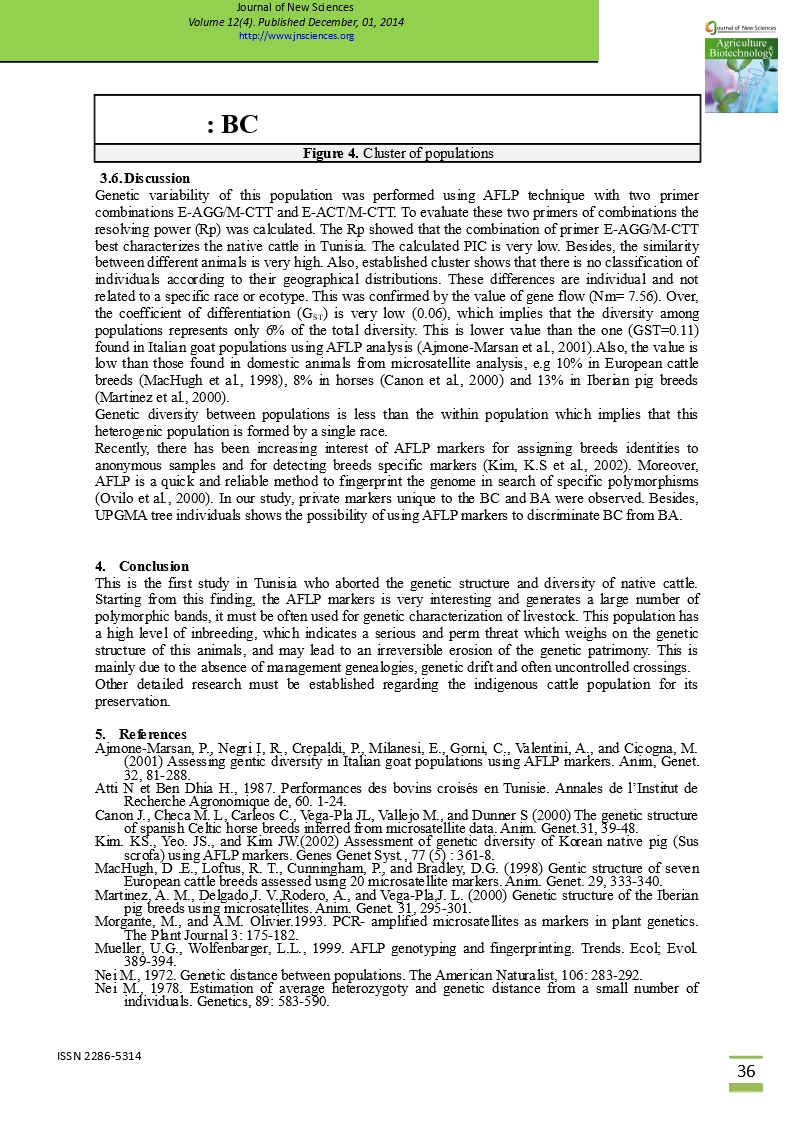
Based on genetic distances, a hierarchical classification was performed (figure 4). It appears that populations 1 and 4 (BAZ and BC) are farthest, while the closest are 1 and 3 (BAZ and BAJ). However, populations 2 and 4 (BAB and BC) are close.
|
|
|
Figure 4. Cluster of populations |

-
Discussion
Genetic variability of this population was performed using AFLP technique with two primer combinations E-AGG/M-CTT and E-ACT/M-CTT. To evaluate these two primers of combinations the resolving power (Rp) was calculated. The Rp showed that the combination of primer E-AGG/M-CTT best characterizes the native cattle in Tunisia. The calculated PIC is very low. Besides, the similarity between different animals is very high. Also, established cluster shows that there is no classification of individuals according to their geographical distributions. These differences are individual and not related to a specific race or ecotype. This was confirmed by the value of gene flow (Nm= 7.56). Over, the coefficient of differentiation (GST) is very low (0.06), which implies that the diversity among populations represents only 6% of the total diversity. This is lower value than the one (GST=0.11) found in Italian goat populations using AFLP analysis (Ajmone-Marsan et al., 2001).Also, the value is low than those found in domestic animals from microsatellite analysis, e.g 10% in European cattle breeds (MacHugh et al., 1998), 8% in horses (Canon et al., 2000) and 13% in Iberian pig breeds (Martinez et al., 2000).
Genetic diversity between populations is less than the within population which implies that this heterogenic population is formed by a single race.
Recently, there has been increasing interest of AFLP markers for assigning breeds identities to anonymous samples and for detecting breeds specific markers (Kim, K.S et al., 2002). Moreover, AFLP is a quick and reliable method to fingerprint the genome in search of specific polymorphisms (Ovilo et al., 2000). In our study, private markers unique to the BC and BA were observed. Besides, UPGMA tree individuals shows the possibility of using AFLP markers to discriminate BC from BA.
-
Conclusion
This is the first study in Tunisia who aborted the genetic structure and diversity of native cattle. Starting from this finding, the AFLP markers is very interesting and generates a large number of polymorphic bands, it must be often used for genetic characterization of livestock. This population has a high level of inbreeding, which indicates a serious and perm threat which weighs on the genetic structure of this animals, and may lead to an irreversible erosion of the genetic patrimony. This is mainly due to the absence of management genealogies, genetic drift and often uncontrolled crossings.
Other detailed research must be established regarding the indigenous cattle population for its preservation.
-
References
Ajmone-Marsan, P., Negri I, R., Crepaldi, P., Milanesi, E., Gorni, C., Valentini, A., and Cicogna, M. (2001) Assessing gentic diversity in Italian goat populations using AFLP markers. Anim, Genet. 32, 81-288.
Atti N et Ben Dhia H., 1987. Performances des bovins croisés en Tunisie. Annales de l’Institut de Recherche Agronomique de, 60. 1-24.
Canon J., Checa M. L, Carleos C., Vega-Pla JL, Vallejo M., and Dunner S (2000) The genetic structure of spanish Celtic horse breeds inferred from microsatellite data. Anim. Genet.31, 39-48.
Kim. KS., Yeo. JS., and Kim JW.(2002) Assessment of genetic diversity of Korean native pig (Sus scrofa) using AFLP markers. Genes Genet Syst., 77 (5) : 361-8.
MacHugh, D .E., Loftus, R. T., Cunningham, P., and Bradley, D.G. (1998) Gentic structure of seven European cattle breeds assessed using 20 microsatellite markers. Anim. Genet. 29, 333-340.
Martinez, A. M., Delgado,J. V.,Rodero, A., and Vega-Pla,J. L. (2000) Genetic structure of the Iberian pig breeds using microsatellites. Anim. Genet. 31, 295-301.
Morgante, M., and A.M. Olivier.1993. PCR- amplified microsatellites as markers in plant genetics. The Plant Journal 3: 175-182.
Mueller, U.G., Wolfenbarger, L.L., 1999. AFLP genotyping and fingerprinting. Trends. Ecol; Evol. 389-394.
Nei M., 1972. Genetic distance between populations. The American Naturalist, 106: 283-292.
Nei M., 1978. Estimation of average heterozygoty and genetic distance from a small number of individuals. Genetics, 89: 583-590.
Nei, M., 1973. Analysis of gene diversity in subdivided populations. Proc. Natl. Acad. Sci. U.S.A. 70, 3321-3323.
Nei, M., Li, W.H., 1979. Mathematical model for studying genetic variation in terms of restriction endonucleases. Proc. Natl. Acad. Sci. U.S.A. 76, 5269-5273.
Ovilo, C, Cervera, M. T., Castellanos, C., and Martinez-Zapater, J.M. (2000) Characterization of Iberian pig genotypes using AFLP markers. Anim. Genet. 31, 117-122.
Powell, W., Morganate, M., Andre, C., Hanafey, M., Vogel, J., Tingey, S., Rafalki, A., 1996. The comparison of RFLP, RAPD, AFLP, and SSR (microsatellite) markers for germplasm analysis. Mol. Breed.2, 255-238.
Prevost, A., Wilkinson, M.J., 1999. Anew system of comparing PCR primers applied to ISSR fingerprinting of potato cultivars. Theor. Appl. Genet. 98, 107-112.
Rohlf F.J., 1998. NTSYSpc. Version 2.10p. Applied Biostatistics, Setauket, NY 11733-2870, USA. http://www.exetersoftware.com/cat/ntsyspc/ntsyspc.html.
Roldan-Ruiz, I., Dendauw, J., VanBockstaele, E., Depicker, A., De Loose, M., 2000. AFLP markers reveal high polymorphic rates in ryegrasses (Lolium spp.). Mol. Breed. 6, 125-134.
Sambrook J., Fritsch E.F et Maniatis T., 1989. Molecular Cloning. A laboratory manual second Edition. Cold Spring Harbor Laboratory Press. New York, USA.
Sokal R.R, Michener C.D, 1958. A statistical method for evaluating systematic relationship. University of Kansas Scientific Bulletin 28, 1409-1438.
Vos P., Hogers R., Bleeker M. et Reijans M., 1995. AFLP: A new technique for DNA fingerprinting. Nucleic Acids (23): 4407-4414.
Xue, J.M., Zhuo, L.H., Zhuo, S.L., 2006 Genetic diversity and geographic pattern of wild lotus (Nelumbo nucifera) in Heilongjiang Province. Chin. Sci. Bull.51, 421-432.
Yeh F.C, Yang R.C, 1999. A joint project POPGENE VERSION1.31.University of Alberta and Tim Boyle, Centre for International Forestry Reasearch ftp //ftp. Microsoft.com/Softlib/MSLFILES/HPGL.EXE.
Marayam Moslemi., Mehdi Zahravi., Gholamreza., and Bakhshi khaniki (2010) Genetic diversity and population genetic structure of pomegranate (Punica granatum L.)in Iran using AFLP markers.
